Gamma Knife treatment is usually done in one session. The treatment process usually starts in the morning of the same day and lasts until noon. If your lesion is large or located near vital brain organs, the number of sessions may be up to 5, depending on the decision of your treatment team.

Ehsan Saraee, MD
Radiation Oncologist
Assisstant Professor of Tehran University of Medical Sciences
Radiosurgery with Gamma Knife ICON device allows physicians to treat various size of tumors in the brain without the need for any kind of surgery. The entire treatment process is non-invasive, and lesions inside the brain are treated by using very precise gamma ray radiation, without the need to perform any surgery.
Brain surgery without any incision!
Contrary to its name, no knife is used at any stage of the treatment. In the treatment of patients with this method, 192 gamma rays are irradiated to the lesion from several angles and are concentrated in a point the size of a tenth of a millimeter (about the size of a sheet of paper). In this method, while a very high dose is irradiated to the brain lesion, the dose to vital organs and normal brain tissue is as low as possible. The non-invasive nature of this treatment will significantly shorten the recovery period.
This level of precision means that the Gamma Knife ICON can target the smallest and most challenging brain tumors or lesions with extreme precision, while protecting the normal brain tissues surrounding the lesion. Gamma Knife radiosurgery is also known as stereotactic radiosurgery and can be used as a very suitable alternative to radiation therapy (radiotherapy) of the whole brain for multiple brain metastases.
Yas Hospital Gamma Knife Center
Gamma Knife ICON under the magnifier!

What diagnoses can be treated?
With the Gamma Knife ICON, a range of diagnoses of brain lesions as small as a few millimeters to as large as a few centimeters can be treated without the need for brain surgery. In this treatment technique, it is possible to treat both malignant and benign lesions.
The following diagnoses are among the most common diagnoses approved for treatment with Gamma Knife:
-
Brain Tumors
-
Brain Metastases
The spread of malignant cells from the place where it was origined to another part of the body is called metastasis. In metastasis, malignant (cancerous) cells break away from the primary tumor, travel through the blood or lymphatic system, and form a new tumor in other organs or tissues of the body. Metastatic tumor is of the same histologic type as the primary tumor. For example, if breast cancer spreads to the lung, the cancer cells that develop in the lung are the same type of breast cancer cells, not the original lung cancer cells. When these cancer cells have limited spread to the brain, there are generally two treatment methods available to physicians. In one of these methods, the patient's entire brain will be irradiated and the malignant cells will be treated wherever they are located in the brain. Among the most important side effects of this treatment method, we can mention memory disorder and patient dysfunction. In patients whose number and volume of brain metastatic lesions are limited, radiosurgery technique using Gamma Knife can be used for their treatment. Among the most important advantages of treating patients with this method, we can point out the very low side effects of the treatment, the possibility of repeating the treatment in new lesions, and the very low dose of radiation for healthy brain tissue. Compared to the stereotactic treatment using the Cyberknife device, the Gamma Knife Icon device has much higher accuracy and much fewer side effects. Also, after using the Gamma Knife device, the radiation dose to healthy brain tissue is much lower than other methods.
-
Acoustic Neuroma/Vestibular Schwannoma
It is one of the most common brain lesions that make up many patients in neurosurgery and radiotherapy departments. One of the most important symptoms of this disease in the early stages is hearing loss. This hearing loss has different degrees in different patients, from very little hearing loss to complete deafness. Vestibular schwannoma or acoustic neuroma is a disease resulting from involvement of the auditory nerve after exiting the brain stem. Simply put, this nerve transmits messages related to hearing and balance to the brain, that's why the involvement of this nerve will cause hearing loss and balance disorder. This lesion does not grow fast by itself, but its proximity to sensitive areas of the brain requires more attention to this lesion. One of the most important organs that is next to this lesion and may be involved in the advanced stages of this disease is the brain stem. Pressure on the brain stem will disrupt several brain mechanisms. The next complication observed in this lesion is paralysis of the facial nerve, which causes many complications for patients (dry eyes, masking of the face). In general, there are three ways to deal with this disease: The first method - Follow-up: One of the treatment methods is to follow up patients whose lesion has a very low growth rate and the size of the lesion is very small. The second method - Gamma Knife Radiosurgery: one of the most accurate treatment methods in all parts of the world is treatment with stereotactic techniques with Gamma Knife device. In this treatment method, the tumor is targeted by gamma rays from dozens of different angles, a high dose of gamma rays is irradiated on it, and its growth is stopped in a high percentage of patients. The success rate of this treatment method in 10 years of follow-up is 95%, which is equivalent to surgery, with the difference that it will have much less complications than surgery. Among the most important complications of surgery, facial nerve paralysis (in 20% of cases) and severe hearing loss (in 30% of cases) can be mentioned. But in the treatment with Gamma Knife, these side effects are 1 to 2% of facial nerve damage (returning to normal in 50% of cases) and about 10 to 20% complete hearing loss. The third method - lesion surgery: in this treatment method, all parts of the lesion are removed, but there is a high probability of complications of facial nerve paralysis or balance disorder in these patients. It should be noted that the decision to use each of the mentioned methods will be made according to the characteristics of the lesion.
-
Meningioma
Meningioma is a type of tumor that develops in the meninges, which are the protective membranes of the brain and spinal cord. These tumors are usually slow-growing and usually non-cancerous, although in rare cases they can be malignant. The exact cause of meningioma is unknown, but it is more commonly diagnosed in older people, especially women. Common symptoms of brain meningioma include headache, seizures, changes in vision or hearing, weakness or numbness in limbs, and cognitive problems. Meningioma treatment options depend on various factors, including tumor size, location, and overall health of the patient. Surgical removal is often the primary treatment, but conventional radiotherapy, Gamma Knife, or targeted drug therapy may also be used to slowen tumor growth. Regular follow-up to monitor tumor progression and ensure early detection of any possible recurrence is of utmost importance in these patients. With proper medical care and support, many patients with cerebral meningioma can lead normal lives.
- Recurrences of Glioblastoma
-
Pituitary adenomas
The pituitary gland is one of the most important secretory glands in the body that controls hormones in your body that have various functions such as stress response, metabolism, growth and sexual function. Pituitary gland tumors can cause a variety of problems. Radiosurgery of the pituitary gland can be used to shrink tumors in this area and reduce hormone regulation disorders.
-
Brain Metastases
-
Vascular lesions
-
Arteriovenous Malformation(AVM)
AVMs are abnormal formations of arteries and veins in your brain. In an AVM, blood flows directly from the arteries into the veins, bypassing smaller blood vessels (capillaries). Over time, AVMs may disrupt normal blood flow and lead to brain hemorrhage or stroke. Stereotactic radiosurgery causes damaged blood vessels to close over time. Of course, it is necessary to mention that in order to observe the treatment response to radiosurgery in AVM patients, a longer period of time is needed compared to angioembolization treatments (injection of blocking substance into the lesion).
-
Arteriovenous Malformation(AVM)
Functional Lesions
- Essential Tremor
-
Trigeminal Neuralgia
Trigeminal Neuralgia is a chronic pain disorder in branches of the trigeminal nerve, which transmits sensory information between the brain and the forehead, cheek, and lower jaw regions. This nerve disorder causes severe pain in the face, which is similar to the application of electric shocks in these areas at time intervals but continuously. Stereotactic radiosurgery treatment for trigeminal neuralgia targets the nerve root to disrupt these pain signals. It should be noted that the selection of patients for Gamma Knife treatment is highly dependent on their response to previous treatments. Somehow, this treatment method is not usually used in the first line of treatment for trigeminal neuralgia patients.
Consult with Ehsan Saraee, MD for the possibility of treatment with Gamma Knife Technique
Saturdays, Mondays, Wednesdays and Thursdays at Yas Hospital Gamma Knife Center
Treatment Steps
In this section, we will explain the steps of treating patients with the Gamma Knife device:
1 - Pre treatment consultation
Before treatment begins, your doctor will discuss your treatment options, explain any side effects you may experience, and answer any questions you may have. It may be better to write down your questions before the consultation and take notes of the information received during the consultation session. During the initial consultation, the consulting physician will check all your documents and after considering your situation, he will inform you of the final decision. Pay attention to the fact that to participate in the counseling session, bring all the patient's documents, including the pathologies and imaging done. Try not to let more than 1 month pass since the last imaging. The presence of the patient is also mandatory in the consultation sessions.
2 - Registering patient
According to the internal rules of the Radiotherapy Department - Gamma Knife Center of Yas Hospital, all patients are required to register in the http://register.gammayas.ir system. After entering your contact number in the system, request a new consultation and upload the patient's history and all the documents related to the disease in the system. Required documents for consultation include your physician's letter of introduction to participate in the scientific committee, pathology reports in case of previous surgery, imaging reports (the most recent imaging should be for the last 1 months) and in patients with functional pituitary adenomas, the latest Blood tests and medications being used. After sending the documents, wait for the response of the system administrator for the final confirmation of receiving the documents. If the submitted documents are adequate, you will be included in the review list of the scientific committee.
3 - Participate in the scientific committee
After the initial review of all patients' documents, if necessary, the patient will be referred to the scientific committee of Yas Hospital Complex. In the Gamma Knife scientific committee, experts in radio-oncology and neurosurgery review all the patients' documents and if the patient is a suitable candidate for treatment with Gamma Knife, they issue the approval for their treatment.
4 - Brain MRI with Gamma Knife protocol
To contour and plan the treatment with the Gamma Knife, within 10 days before the start of the treatment, you must go to one of the MRI centers introduced by Gamma Knife responsible staff and perform the requested MRI imaging. After the imaging, you will deliver the CD containing the relevant images to the Gamma Knife department, and the colleagues will enter all the information of the received images into the treatment planning software(TPS). At this time, a medical file will also be created for you in the software.
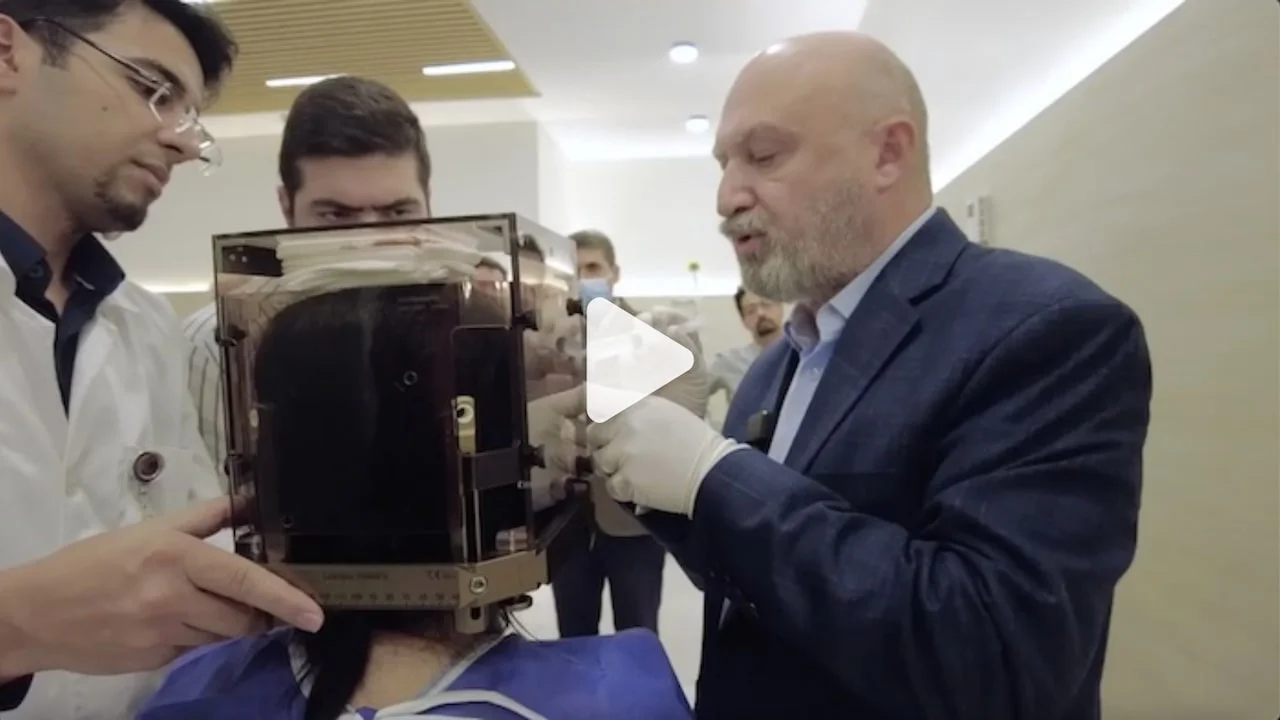
5 - Stabilization of head position during treatment
To achieve the highest precision in the treatment process, it is important that your head is held in place with a custom thermoplastic face mask or a metal frame.
If your treatment team decides to treat you in one session, they will install a titanium frame on your head with four special pins. This frame will stabilize your head during the several-hour treatment period and will serve as a reference point for focusing the radiation beams. During the insertion of the metal frame, local anesthetics will be injected into the four points on your scalp where the pins will be placed (two on the forehead and two on the back of the head). In most cases, you will not need to shave your head. Some types of brain radiosurgery may not require the installation of a metal frame on the head, and the treatment of patients will be done through the installation of special masks. If you are treated using a mask, this mask will be custom-made for you in the department before the start of your treatment process. The mentioned mask is made of lightweight material with small holes. After placing the mask in water with a certain temperature, it becomes flexible and meanwhile, it is placed on your face and after about 10 minutes, it will completely find the shape of your head and face. After the complete preparation of the desired mask, your head will be kept in its position with great precision during the Gamma Knife treatment. Meanwhile, the camera monitors the position of your head during the treatment and sends a report of any movement in the position of the head to the treatment planning software.
6 - Radiosurgery Planning
After entering your imaging information into the treatment planning software, your treatment team will start the contouring process. In this process, in each of the MRI slices taken, the extent of the brain lesion is determined accurately, and then the center's physicist will plan your treatment according to this information.
7 - Treatment
When it's time for your treatment, the treatment team will help you get on the treatment bed. If your treatment is with a mask, at this stage the mask will be placed on your face. A marker may be placed on the tip of your nose. A camera uses this indicator to monitor any movement of your head during treatment. Before starting the treatment, a CT scan of your brain is performed and the resulting images are sent to the treatment planning software, and using this imaging information, the exact position of your mass is calculated in relation to the device. Depending on the size, shape and complexity of the brain tumors being treated, the treatment may last from a few minutes to over an hour. You will not feel anything during the treatment. In fact, some patients even fall asleep. You can communicate with your treatment team at any time through the device's internal intercom.
8 - Follow up
After treatment, you can usually go back to your normal activities in a day or so. You will see the effect of treatment with this method usually in weeks or months after the treatment. The EMR designed by me in Yas Hospital will automatically send an SMS to the number registered in the system at the appropriate time for your follow-up. You may need to do a new MRI, audiometry, or vision tests in your follow-up visits.
In what conditions is Gamma Knife treatment recommended?
If a tumor or lesion is located near one of the dangerous areas in the brain, stereotactic Gamma Knife radiosurgery may be recommended. This part can be the area responsible for important functions such as speech or movement.
When tumors or lesions are located deep in the brain, in some situations the Gamma Knife is a safer option than open brain surgery, because reaching that area can be very dangerous depending on whether there are blood vessels or other vital areas. Also, this method is a low-risk solution for patients who do not have suitable conditions for brain surgery or are at high risks of anesthesia and cardiac complications, which means that surgery is not suitable for them.
Also, Gamma Knife radiosurgery is an effective treatment option for multiple brain metastases. The low dose reached to healthy brain tissue has the advantage that this treatment can be repeated for patients if needed. This means that whole brain radiotherapy can be avoided or delayed in many patients with brain metastases. It should be noted that this treatment method is not suitable for all patients and should be done according to the discretion of the treatment team and the scientific committee.
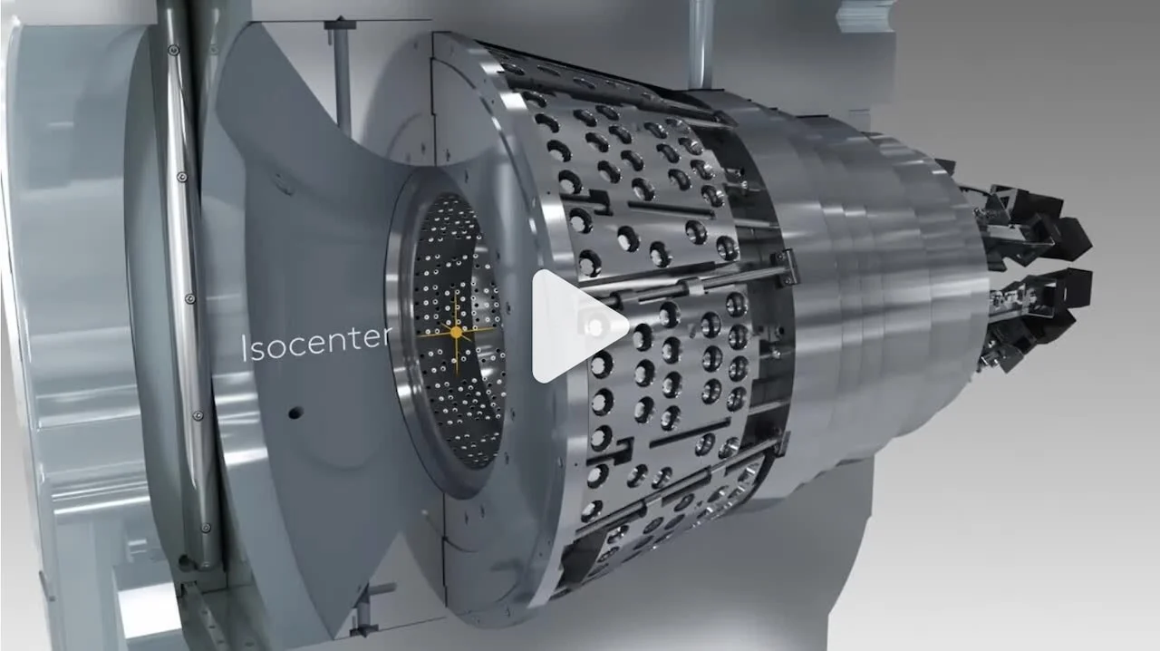
What does Gamma Knife look like?
How does Gamma Knife work?
Gamma Knife treats brain lesions by irradiating gamma rays completely focused on the lesion, so that it is possible for the treated lesion to disappear, shrink, or stop growing. This treatment method can be used for lesions in the most critical and inaccessible areas of the brain without sending significant doses of radiation to healthy brain tissue.
Can Gamma Knife be used to treat lesions of the whole body?
Gamma Knife is specifically designed for precise treatments in the brain and is capable of treating very complex and challenging lesions or tumors in all parts of the brain. This treatment method cannot be used for lesions outside the brain.
How many days does Gamma Knife treatment take?
In most cases, the treatment is done in one session and on an outpatient basis, patients usually receive their treatment plan including the number of treatment days from their treatment team on the first day of visit. The treatment time in each session can be from a few minutes to an hour or more. In a group of patients, due to the size of their lesion, the same procedure may take place within a few days. Patients can usually resume their normal activities shortly after treatment.
What is the difference between Gamma Knife and other stereotactic treatment methods such as Cyberknife?
2-4X
Lower dose reached the normal brain tissue
In general, Gamma Knife has a higher ability to protect normal brain tissue from radiation.
2-21X
The dose reached the whole body
Compared to other brain tumor treatments
Up to 3X
Further clinical studies
Compared to other medical devices
The most common questions about Gamma Knife:
Gamma Knife treatment is usually done in how many sessions?
Will I feel anything during the Gamma Knife treatment?
Normally, you will not feel anything during the treatment. If your treatment requires a frame to be placed on the head, you may feel a slight pain from the administration of the local anesthetic used during the placement of the frame. A group of patients reported feeling pain or pressure during the installation of the frame. It should be noted that, in most cases, the implanted frame will be removed from the patient's head in less than an hour.
What side effects can I expect after treatment with Gamma Knife?
After the end of the treatment session, the frame installed on the head or the mask placed on the face is removed. Sometimes a little bleeding will occur from the contact point of the pins with the head, which in many cases can be controlled by just applying pressure to the area. It is usually recommended that the patient refrain from vigorous physical activity for the next 18 to 24 hours, but after that he can resume his daily activities.
How long will it take to see a therapeutic response after treatment with Gamma Knife?
Depending on the type of lesion treated, the speed of response to treatment will be different. For example, in patients with brain metastases, response to treatment is usually observed one to two months after treatment. On the other hand, patients with vestibular schwannoma, a therapeutic response with be achievable in more than one year. In patients with functional pituitary adenomas, hormones may return to normal levels after one to two years.
How many times can you do Gamma Knife?
Yes, due to the very low dose reaching the normal brain tissue, this treatment method can be repeated several times.
Get more information
You can consult with us for a more detailed review of the case and to answer your questions regarding treatment with Gamma Knife.
Saturdays, Mondays, Wednesdays and Thursdays at Gamma Knife Center of Yas Hospital